
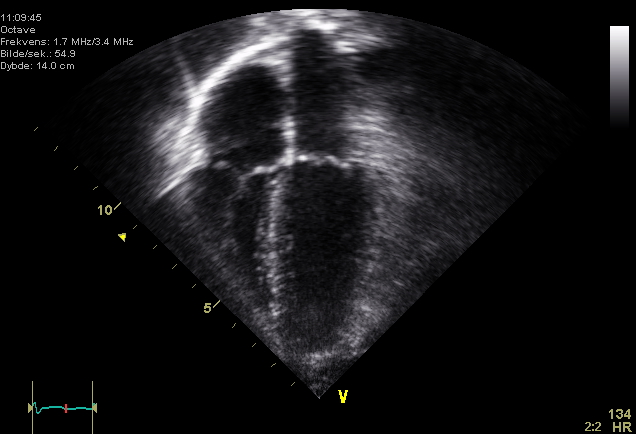
Echocardiogram
An echocardiogram is involves producing an image of your heart via use of soundwaves. The picture is highly detailed and does not involve exposure to ionizing radiation.
How is the test is carried out?
A trained sonographer performs the test, then your heart doctor interprets the results. High-frequency sound waves from a transducer placed on the ribs are directed toward the heart.
Other images are obtained from underneath and slightly to the left of your nipple (at the apex of your heart). The transducer picks up the echoes of the sound waves and transmits them as electrical impulses. Pictures can then be produced from these electrical impulses by the echocaridography machine.
Resources
Image courtesy of Wikki Commons
https://commons.wikimedia.org/wiki/File:Echocardiogram_4chambers.jpg